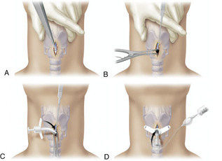
This week in lab we were dissecting the muscles of the face and examining the structures of the neck. Before I get to talking about the cricothyrotomy, lemme just say, the facial muscles are so superficial. Trying to reveal them was like basically shaving just a hair deeper. This is probably why when people have face lacerations that we need to use such a fine suture and call the plastic surgeon instead of having the ER docs just sew it up. Unfortunately we only could recover some of the facial muscles to look as pretty as we wanted them to. Our cadaver had lots of facial hair so it was sad to cut it off. I'm sure he was the kinda guy who wouldn't shave for anything (like most men)! We revealed the submandibular glands, and boy were they much larger than I expected! We also cleared away enough to see the thyroid and the cricoid cartilage. Now, I was not able to do a cricothyrotomy myself because someone else in our group was all over trying it themselves. (remember that each person only has one neck, so its not like everyone can do a cricothyrotomy on all the cadavers since we only have 5). One of our group-mates was very enthusiastic about it and tried it on a cadaver. So basically what she did is in the picture below. She punctured the space just above the cricoid cartilage to open the airway. This would be necessary if someone had a blockage above or could not breathe. Since we wanted to get a better look at the internal structures, we used a bone saw to open up the cartilage and reveal the true and false vocal cords. Overall, it was pretty cool. However, we also got a great lecture from one of the doctors in our lab. He went through a detailed lecture on the sagittal section of the head and neck for us to be able to see what was inside.
This was probably the coolest thing I saw so far. Below is an example of what the sagittal section would look like on a model. We were able to see the brain (literally that comparison of tapioca pudding is 100% accurate), the conchae of the nose, the structures of the neck, all the sinuses and so much more. I did not realize what help this cadaver lab would be to us, but it sure helps me remember so much!
This was probably the coolest thing I saw so far. Below is an example of what the sagittal section would look like on a model. We were able to see the brain (literally that comparison of tapioca pudding is 100% accurate), the conchae of the nose, the structures of the neck, all the sinuses and so much more. I did not realize what help this cadaver lab would be to us, but it sure helps me remember so much!
 |  |