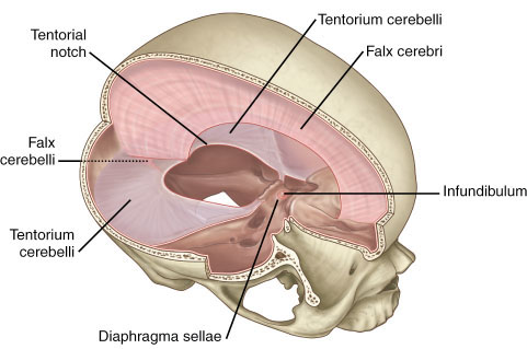
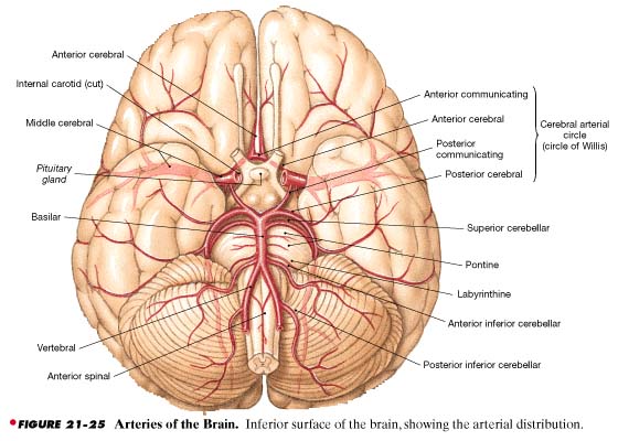
Our last official anatomy lab involved exploration of the brain. We were warned ahead of time that the brains may be very squishy due to the enbombing; however, our cadaver's brain was well preserved. First, since our cadaver had a full head of hair, we had to scalp off his hair and his skin to get to the bone. Then, we used the same bone saw that we used in the second week of lab (for the laminectomy) for the calvaria removal. Unlike the spine, the use of the bone saw on the skull requires a bit more talent because it requires you to carefully trace around the head without making a curved jagged cut. The thickest part of skull is the occiput where we needed a little more elbow grease in order to get the saw through. Now, even though we cut the bone, the calvaria did not easily pop off. The falx cerebri is an attachment between the brain and the bone beneath the sagittal suture (you can see this in the image below). Also, the cerebellum is contained below the tentorium cerebrelli which means we needed to carefully trace out the brain edge of it. Just before the brain was released from the skull, it was connected by the cranial nerves, blood vessels and the spinal cord. Once the these were cut, the brain came right out of the skull. Our view was that of the second picture below. Here we could view all the vessels. However, the best part was that our cadaver had an extra blood vessel coming from his middle cerebral artery leading to the posterior communicating (maybe even the posterior cerebral). Our professors were surprised and actually have never seen this in someone before so they had to look up some anomalies to see how common it is.
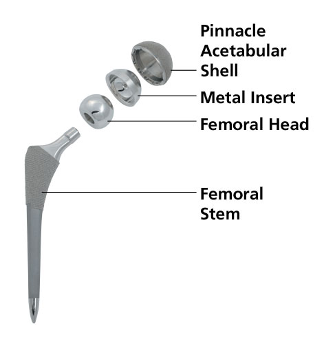
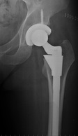
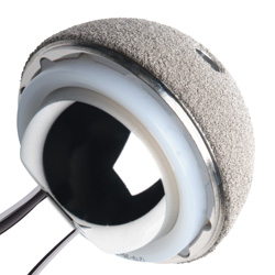
Aside from the brain our cadaver was also dissected more from the hip because there were large visible staples that may have been evidence of a recent surgery. After chiseling away, our classmates found a shiny steel hip replacement. They were able to clear away the bone on the femur and around the acetabulum in order to get a better look at the prosthetic. The third picture below is what the implant looked like.
Aside from the brain our cadaver was also dissected more from the hip because there were large visible staples that may have been evidence of a recent surgery. After chiseling away, our classmates found a shiny steel hip replacement. They were able to clear away the bone on the femur and around the acetabulum in order to get a better look at the prosthetic. The third picture below is what the implant looked like.
 |  |  |